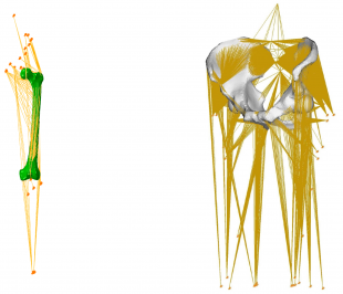
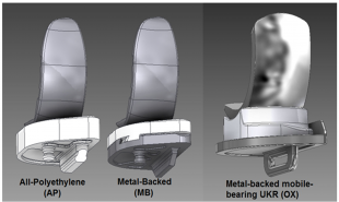
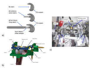
We have developed one of the most biofidelic numerical models of the human pelvis, which for the first time included muscular and ligamentous supports [1]. Complex nonlinear models were developed to examine migration of implants after hip replacement (observed clinically) [2]. This work won the best medical engineering PhD award (2005) from the Institution of Mechanical Engineers (IMechE). Ongoing research focuses on implant instability and failures after total [3-5] and partial knee replacements [6-7]. A biomechanical study on unilateral knee replacements received a number of awards including best medical engineering project prize from the IMechE.
References:
- A.T.M. Phillips, P. Pankaj, C.R. Howie, A.S. Usmani, A.H.R.W. Simpson, Finite element modelling of the pelvis: inclusion of muscular and ligamentous boundary conditions. Medical Engineering & Physics, 29 (2007), 739–748. DOI:
10.1016/j.medengphy.2006.08.010 - A.T.M. Phillips, P. Pankaj, C.R. Howie, A.S. Usmani, A.H.R.W. Simpson, 3D non-linear analysis of the acetabular construct following impaction grafting. Computer Methods in Biomechanics and Biomedical Engineering, 9 (2006), 125-133. DOI:10.1080/10255840600732226
- N Conlisk, H Gray, P Pankaj, CR Howie, The influence of stem length and fixation on initial femoral component stability in revision total knee replacement. Bone and Joint Research 1 (11), 281-288. DOI:
- N Conlisk, H Gray, P Pankaj, CR Howie, The role of complex clinical scenarios in the failure of modular components following revision total knee arthroplasty: A finite element study. Journal of Orthopaedic Research 33 (8), 1134-1141. DOI:10.1002/jor.22894
- N Conlisk, H Gray, P Pankaj, CR Howie, An efficient method to capture the impact of total knee replacement on a variety of simulated patient types: A finite element study. Medical Engineering & Physics 38 (9), 959-968. DOI:http://dx.doi.org/10.1016/j.medengphy.2016.06.014
- Scott, CEH, Eaton, MJ, Wade, FA, Nutton, RW, Evans, SL & Pankaj, P 2016, 'Metal backed versus all-polyethylene unicompartmental knee arthroplasty: the effect of implant thickness on proximal tibial strain in an experimentally validated finite element model' Bone & Joint Research.
- Scott, CEH, Wade, FA, Bhattacharya, R, MacDonald, D, Pankaj, P & Nutton, RW 2016, 'Changes in Bone Density in Metal-Backed and All-Polyethylene Medial Unicompartmental Knee Arthroplasty' Journal of Arthroplasty, vol 31, no. 3, pp. 702–709., DOI:10.1016/j.arth.2015.09.046


