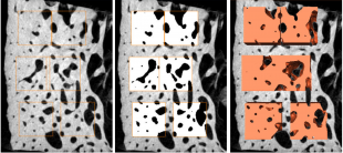
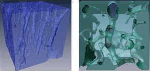
Our research has shown for the first time that cortical bone becomes more anisotropic with age [1], which results in increased vulnerability to fracture due to forces in the less frequently loaded directions. We have developed novel nonlinear homogenisation approaches to determine the post-elastic macro-mechanical behaviour of bone from its microstructure [2]. This research will will enable diagnosis of bone quality from its images and permit realistic evaluation of bone and bone-implant systems. Our bone micromechanics research has found an application in the evaluation of snow properties for avalanche prediction [3].
References:
- Donaldson, FE, Pankaj, P, Cooper, DML, Thomas, CDL, Clement, JG & Simpson, H 2011, 'Relating age and micro-architecture with apparent-level elastic constants: An FE study of female cortical bone from the anterior femoral midshaft' Proceedings of the Institution of Mechanical Engineers, Part H: Journal of Engineering in Medicine, vol 225, pp. 585-596. DOI:10.1177/2041303310395675
- Levrero-Florencio, F, Margetts, L, Sales, E, Xie, S, Manda, K & Pankaj, P 2016, 'Evaluating the macroscopic yield behaviour of trabecular bone using a nonlinear homogenisation approach' Journal of the mechanical behavior of biomedical materials, vol 61, pp. 384-396., DOI:10.1016/j.jmbbm.2016.04.008
- Srivastava, PK, Chandel, C, Mahajan, P & Pankaj, P 2016, 'Prediction of anisotropic elastic properties of snow from its microstructure' Cold Regions Science and Technology, vol 125, pp. 85-100., DOI:10.1016/j.coldregions.2016.02.002


